
價格:免費
更新日期:2012-07-30
檔案大小:14M
目前版本:1.2
版本需求:Android 2.2 以上版本
官方網站:http://www.libroscience.com
Email:admin@libroscience.com
聯絡地址:KDDI Blding Annex 2F 2-3-3 Nishi-Shinjuku, Shinjuku-ku Tokyo 160-0023 JAPAN

★Lite version★
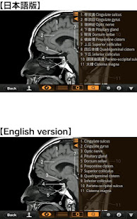
This is the free Lite version of "Interactive CT and MRI Anatomy".
The function is restricted.
You can only see the transverse CT images of the head.
Please check the operation before purchasing the full version.
★ Details ★
This application is developed for medical students, interns, residents, doctors, nurses, and radiology technicians to understand the essential anatomical terms of the body.
You can learn anatomy by answering the terms by step-to-step questions using a total of 241 CT and MRI images.
A total of 17 images of 3D-CT, MRA and plain X-ray film(particularly the extremities) are included as references.
Other reference images include coronary artery segments defined by the American Heart Association(AHA), pulmonary segments, and liver segments(according to Couinaud classification).
You can enlarge all the images by simple manipulation.
★ Major functions ★
There are 4 major functions.
-1) Anatomical mode

Anatomical terms are overlaid on the images.
It can be used as the anatomical atlas.
-2) Quiz mode type 1
You select the part of the image by using anatomical term.
Questions will basically appear randomly.
-3) Quiz mode type 2
You select the anatomical term by the part of the image.
Questions will basically appear randomly.
-4) Index
You can find the specific images by using anatomical terms.
★ Intended users ★
-Medical students
-Interns and residents
-Doctrors
-Nurses
-Radiology technicians
-All those who are intrested in CT and MRI anatomy
★ Images(a total of 258 images) ★
Images basically include horizontal, coronal, and sagital planes.
-Head(36 images including CTA and 3D-CT)
-Neck(24 images)
-Spine(19 images including plain X-ray films)
-Chest(61 images including 3D-CT images)
-Abdomen (37 images)
-Pelves: male (9 images)
-Pelvis: female (11 images)
-Extremities (shoulder, hand, elbow, hip joint, knee, foot) (61 images including plain X-ray films)
Editors

Toshiaki Nitori, M.D. (Professor of Radiology, Kyorin University, School of Medicine)
Yasuo Sasaki, M.D. (Manager of diagnostic radiology, Iwate Prefectural Central Hospital)
